Figure 2.

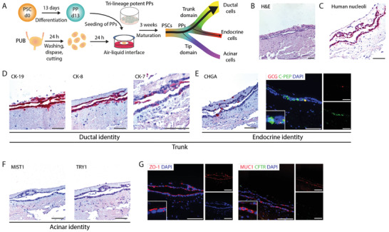
Pancreatic progenitors differentiate towards trunk lineage on the porcine urinary bladder. A) Schematic illustration of the culture of pancreatic progenitor (PP) cells on de‐epithelialized porcine urinary bladder. B) Hematoxylin and eosin (H&E) stained histological sections of PPs cultured on porcine urinary bladder (n = 2). Immunohistochemistry stainings for C) human nucleoli and D) duct‐specific cytokeratins CK‐19, CK‐8, and CK‐7 on PPs cultured on porcine urinary bladder (n = 2). E) Immunohistochemistry stainings for endocrine‐specific chromogranin A (CHGA) and immunofluorescence stainings for C‐peptide (C‐PEP, green) and glucagon (GCG, red) on PPs cultured on porcine urinary bladder (n = 2). Cells were counterstained with DAPI (blue). F) Immunohistochemistry stainings of acinar‐specific markers MIST1 and TRY1 on PPs cultured on porcine urinary bladder (n = 2). G) Immunofluorescence stainings for ZO‐1 (red) and for MUC1 (red) and CFTR (green) (n = 2). Cells were counterstained with DAPI (blue). Scale bars represent 100 µm. PSC, pluripotent stem cells; PUB, porcine urinary bladder.
