Figure 8.

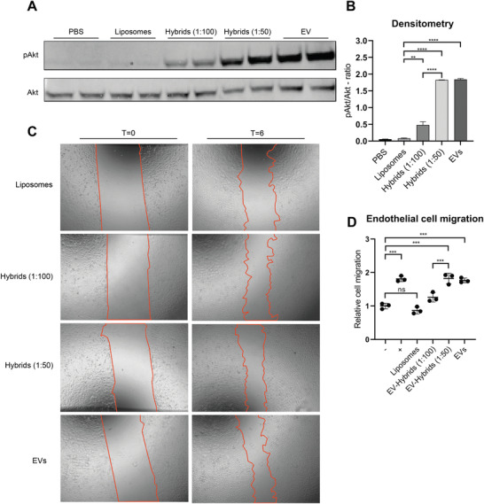
Endothelial signaling assay and scratch wound closing assay of HMEC‐1 cells treated with liposomes and hybrids. A) Representative western blot analysis of phosphorylated Akt and Akt expression levels in HMEC‐1 cells treated with liposomes, hybrids, and EVs. Liposomes and hybrids were administered at a total particle dose of 2 × 10¹² and EVs at a total particle dose of 3 × 10¹⁰. B) Quantification of Akt and pAkt expression levels obtained via western blot analysis using densitometry expressed as pAkt/Akt‐ratio. C) Representative images of scratch wound healing assay before (t = 0) and after (t = 6 h) incubation with liposomes, hybrids (1:100), hybrids (1:50), and EVs. Liposomes and hybrids were administered at a total particle dose of 2 × 10¹² and EVs at a total particle dose of 3 × 10¹⁰. D) Cell migration of HMEC‐1 expressed relative to the negative control. Incubation of HMEC‐1 cells with hybrids (1:50 and 1:100) and EVs increases wound closure as compared to liposomes. Data are expressed as mean + SD, one‐way ANOVA with post‐hoc test, ns = not significant, ** p < 0.01, *** p < 0.001, and **** p < 0.0001.
