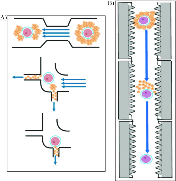
Fig. 2.
The denudation devices proposed by: (A) Zeringue et al. (2001) and (B) Weng et al. (2018)
(A) Device created from polydimethylsiloxane with channels of oocyte-cumulus complex size (400 μm) is implemented for tracking and denudation of the oocytes. Two narrow regions of the microchip reorient the cumulus into a doughnut shape, while two ports (channels significantly narrower than the ovum) are used to remove the cumulus. The device uses pressure-driven flow to properly position and denude the oocytes. (B) The microfluidic device implicates denudation of enzyme-treated cumulus–oocyte complexes by flow-guided passage through a series of jagged-surface constriction microchannels of optimized geometries. The jagged inner wall of the channels is stripping off cumulus–corona cell mass. Reprint with permission from “Automation in Clinical Embryology Laboratories – What is Next?” by Halicigil C., Ogut M.G., Demirci U. © 2023, World Scientific

