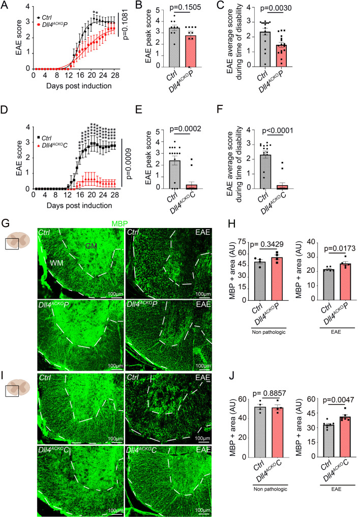
Fig. 2.
Inactivation of astrocytic Dll4 reduces disability during the onset and plateau of EAE disease. (A) Dll4ACKOP mice and control mice induced with EAE were scored daily according to a widely-used 5-point scale (EAE scoring: 1 limp tail; 2 limp tail and weakness of hind limb; 3 limp tail and complete paralysis of hind legs; 4 limp tail, complete hind leg and partial front leg paralysis). Statistical significance was determined by using a 2 ways Anova test followed by the Holm-Sidak’s multiple comparisons test (*: p ≤ 0.05; **: p ≤ 0.01; ***: p ≤ 0.001 ****: p ≤ 0.0001). (Dll4ACKOP n = 10, WT n = 9). (B) Dll4ACKOP and control mice EAE peak score and (C) EAE average score during time of disability (mice present symptoms) were quantified other the course of the disease. (D) Dll4ACKOC mice and control mice induced with EAE were scored daily on a standard 5-point scale, two ways Anova. (Dll4ACKOC n = 15, WT n = 15). (E) Dll4ACKOC and control mice EAE peak score and (F) EAE average score during time of disability were quantified other the course of the disease. (G-J) Non-pathologic spinal cord tissues and spinal cord EAE lesions from Dll4ACKOP mice, Dll4ACKOC mice and littermate controls were harvested at 18 days post induction. (G-H) Dll4ACKOP mice and control lesions (low magnification images) and (I-J) Dll4ACKOC mice and control lesions (low magnification images) were immuno-stained with an anti-MBP (in green) antibody. (H, J) White matter MBP positive areas were quantified (the white matter (WM) is delimited from the grey matter (GM) by the white dotted lines). (H) (Non-pathologic tissues: Dll4ACKOP mice n = 4, WT n = 4; EAE tissues: Dll4ACKOP mice n = 5, WT n = 6), (J) (Non-pathologic tissues: Dll4ACKOC mice n = 4, WT n = 4; EAE tissues: Dll4ACKOC mice n = 6, WT n = 7)). Statistical significance was determined by using a Mann-Whitney U test

