Figure 4.

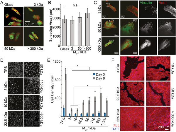
Stem cell expansion at liquid interfaces correlates with interfacial toughness. A,B) Impact of PLL molecular weight on cell spreading at Novec 7500 interfaces stabilized by corresponding nanosheets. C) Confocal microscopy images of MSCs spreading (after 24 h) on PLL/FN functionalized Novec 7500 interfaces. Zoom‐in correspond to the dotted boxes. D,E) MSC expansion at PLL‐stabilized Novec 7500 interfaces (D, representative nuclear stainings). F) Highly confluent MSCs remodel and fracture PLL/FN nanosheets assembled at the surface of Novec 7500. Epifluorescence microscopy images of PLL nanosheets 24 h after seeding MSCs at 200 000 cell/well (left). Red, PLL; blue, nuclei. Detail of interfaces: Novec 7500 containing 10 µg mL−1 PFBC; aqueous solution is PBS with pH adjusted to 10.5; PLL with different M w (3, 10, 22.5, 50, 110, 225, and >300 kDa) at a final concentration of 100 µg mL−1. Error bars are s.e.m.; n ≥ 4. One‐way ANOVA; n.s., non‐significant; *p < 0.05.
