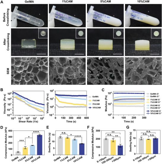
Figure 4.

Characterization of the bioink with different CAM concentrations and particle sizes. A) Gross appearances of bioinks before and after crosslinking, and SEM images of the crosslinked GelMA bioinks containing 0%, 1%, 5% and 10% CAM (scale bar: 200 µm). B) Viscosities of preprinted bioinks and C) storage (G′) and loss (G″) modulus of the bioinks measured by rheological testing. D) Compressive modulus and E) swelling ratio of bioinks containing different CAM concentrations (*p < 0.05, ***p < 0.001, ****p < 0.0001). F) Compressive modulus and G) swelling ratio of bioinks containing 10% w/v CAM of different particle sizes (**p < 0.01).
