Figure 8.

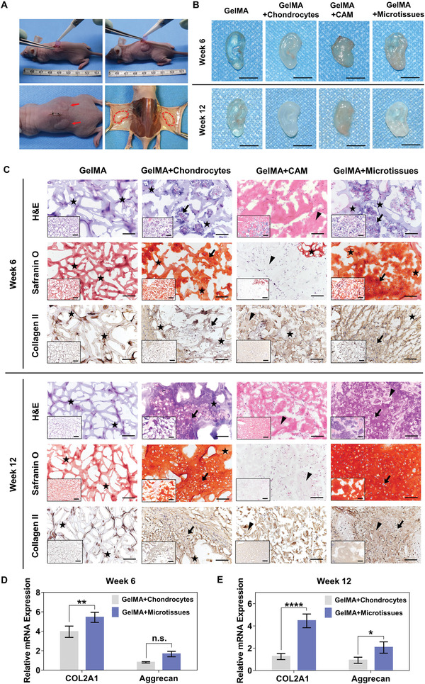
In vivo neocartilage formation. A) Top: the procedure of subcutaneous implantation with auricular constructs in nude mice. Bottom‐left: picture taken right after the subcutaneous implantation of auricular constructs. Bottom‐right: picture taken during explantation of constructs after 12 weeks in vivo. B) Pictures of in vivo constructs at week6 and week12 (scale bar: 1 mm). C) In vivo histological analysis for development of cartilage tissue formation at 6 and 12 weeks after transplantation. Histological and immunohistological stainings for H&E, Safranin O and Collagen II at low magnification (bottom‐left of each picture, scale bar: 500 µm) and at high magnification (scale bar: 100 µm). Asterisk indicates the hydrogel component remaining in the grafts. Triangular arrowhead indicates residual CAM microparticles in the graft. Bold arrow indicates chondrocytes sit in lacunae and are surrounded by the matrix they have secreted. Compared with other three groups, more chondrocytes, red‐stained aggrecan and brown‐stained collagen fibers were observed in GelMA+microtissues group, especially at week12. D,E) mRNA expression analysis of chondrocytes in auricular constructs at week 6 and week 12 (*p < 0.05, **p < 0.01, ****p < 0.0001).
