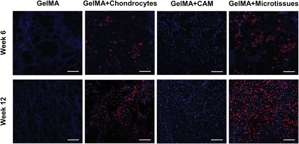
Figure 9.

Immunofluorescence staining images of anti‐human LaminA/C in subcutaneous auricular constructs at week6 and week12. A large number of positive cells were found in GelMA+chondrocytes and GelMA+microtissues groups, indicating that most of the proliferating chondrocytes were derived from human sources, while only blue‐stained nuclei were found in the GelMA+CAM group, indicating that the proliferating cells were derived from mice. In the GelMA group, only blue‐stained hydrogel components were visible, with almost no nuclear components (scale bar: 200 µm).
