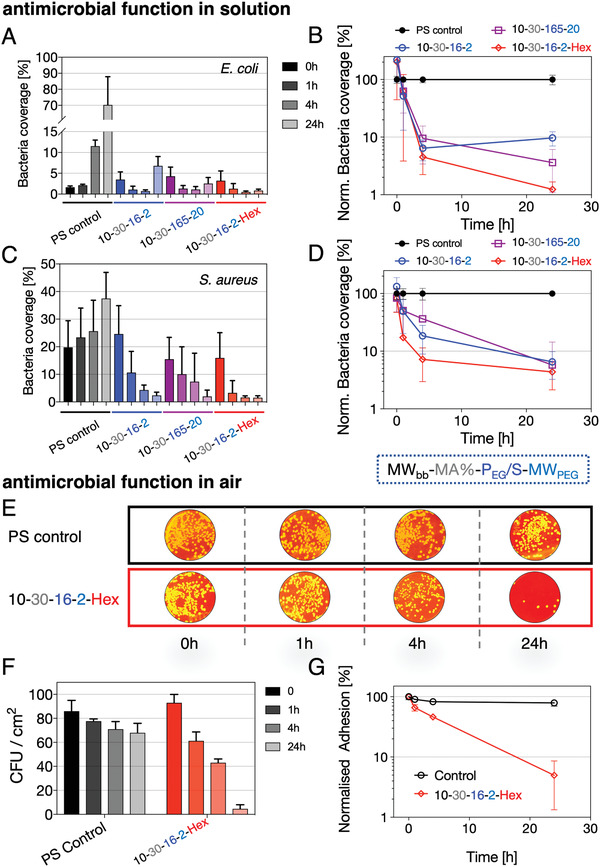
Figure 3.

Antibioadhesiveness and antimicrobial functionality of AP layers. Solution‐based assay of E. coli and S. aureus in contact with antiadhesive 10–30–16–2 and 10–30–165 as well as antiadhesive and antimicrobial 10–30–16–2–Hex (hexetidine) AP layers. A,C) Relative bacterial coverage (100% coverage corresponds to completely covered substrate) over 24 h and B,D) normalized (to control surface) bacteria coverage over time for E. coli and S. aureus. E) S. aureus solutions are directly applied to the substrates and the number of colony‐forming units (CFU) is analyzed after pressing the substrates directly on agar plates (false‐colored photographic images of CFUs on agar plates for better visualization) after the indicated incubation times of 0, 1, 4, and 24 h (gray: control surface, green: 10–30–16–2–Hex). F) Analysis of CFU per cm2 for direct‐contact assay. G) Normalized adhesion of CFUs from (F) indicating the relative reduction in adhered colonies over a time period of 24 h compared to the starting condition.
