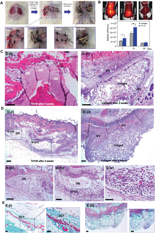
Figure 3.

Soft‐tissue healing and angioconductive properties of TX140. A) Intraoperative wounds after harvesting full‐thickness skin grafts and grafting, treated with TX140 (left) and commercially available collagen scaffold as the gold standard (right) at day 0 and day 7 post skin grafting (n = 27). B) Radiant efficiency of TX140 and collagen scaffold in a murine model after 7, 14, and 28 days (n = 6, statistical significance * for p < 0.05 and ** for p < 0.01)). C) H&E stained cross‐sections of TX140 i) and collagen scaffold ii) after 14 days. D) H&E stained cross‐sections of TX140 i) and the collagen scaffold ii) after 28 days. Cross sections of TX140 iii–v). E) MT stained cross‐sections of Integra and TX140 i) and collagen scaffold ii) biopsies 28 days postskin grafting. GR: TX140 hydrogel residues and IFV: inflammatory fibrosis. DCT: dermal connective tissue. The scale bar in all panel is 100 µm. For (C)–(E) n = 27 in total and 12 at each time point.
