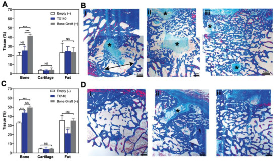
Figure 4.

Hard‐tissue healing in sheep osteotomy model. A) Bone, cartilage, and fat formation in osteotomy sheep model after 6 weeks. B) Masson's Trichrome (MT)‐stained decalcified sections of lesion explants from i) Empty, ii) TX140, and iii) bone graft groups after 6‐weeks postsurgery. C) Bone, cartilage, and fat formation in an osteotomy sheep model after 12 weeks postsurgery D) MT‐stained decalcified sections of lesion explants from i) Empty, ii) TX140, and iii) bone graft groups after 12‐weeks postsurgery. The scale bar in all panels is 1 mm. Asterisks highlight the new bone formation foci of fibrous connective tissue (n = 6).
