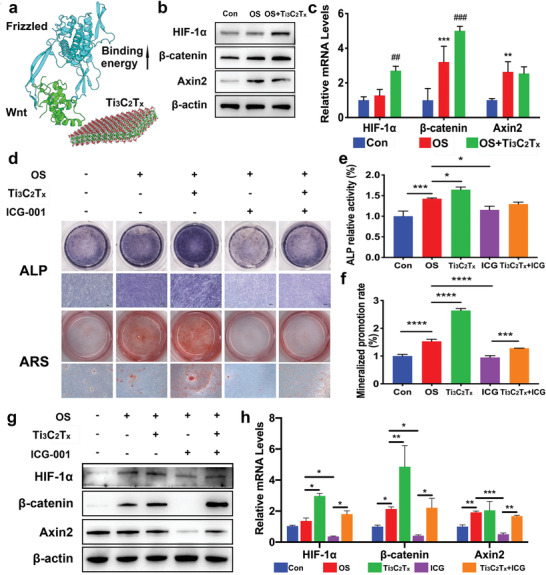
Figure 4.

Effects of the HIF‐1α/WNT signaling pathway on osteogenic differentiation of hPDLCs induced by Ti3C2T x . hPDLCs were treated with Ti3C2T x (60 mg L−1). a) Binding energy difference estimated in the presence of Ti3C2T x in the vicinity of the Wnt‐Frizzled complex. b) Relative protein levels of HIF‐1α, β‐catenin, and Axin2 determined by western blots on day 7. c) The mRNA expression of HIF‐1α, β‐catenin, and Axin2 on day 5 analyzed by real‐time PCR. For analysis of the role of the WNT/β‐catenin signaling pathway in the osteogenic differentiation promoted by Ti3C2T x , hPDLCs were incubated with ICG‐001 (10 × 10−6 m). d) ALP staining on day 7 and mineralized nodules stained with ARS on day 21. e) ALP activity levels on day 7. f) Mineralized nodule levels on day 21. ICG‐001 blocked the protein expression of HIF‐1α, β‐catenin, and Axin2 on day 7 g) and the mRNA expression h) on day 5. *P < 0.05, **P < 0.01, ***P < 0.001, ****P < 0.0001, compared with the Con group or the corresponding group. ##P < 0.01, ###P < 0.001, compared with the PDLC group.
