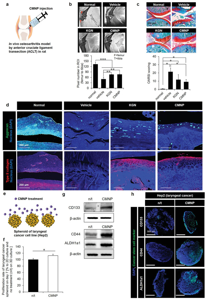
Figure 2.

Chondroprotection and cancer survival due to the prohypoxic effect of CMNPs as a promoter of cell–cell interaction. a) After 6 weeks of induction of ACLT osteoarthritis in rats, CMNP treatment promoted cartilage reinforcement to the level obtained with KGN treatment for the next 6 weeks (of the 12 weeks total experimental period). b) This was evidenced by (top) the end‐point MRI images of sagittal knee joints and (bottom) quantitative analysis of pixel numbers in the ROI from the femur to the tibia using ImageJ (F = femur, T = tibia). c) Safranin‐O staining confirmed the 12‐week results: (top) the histology images (red = cartilage, blue = bone, white = synovium) and (bottom) OARSI scores from the analysis of medial tibial plateaus (F = femur, T = tibia), as supported by d) the immunohistochemistry images of aggrecan (green) and type II collagen (red). e) Induction of inner spheroid hypoxia by 7‐day treatment of CMNPs (20 µg mL−1) promoted f) laryngeal cancer cell (Hep2) proliferation with increased protein expression of cancer stem cell markers (CD133, CD44, and ALDH1a1) according to g) western blotting and h) immunocytochemistry analyses. The proliferation rate of Hep2 cells in 3D hypoxic culture was normalized to that of 2D nonhypoxic culture to exclude the effects of unidentified constituents inside CMNPs, thereby capturing only the effect of hypoxia. Data are expressed as mean ± standard deviation (SD). *p < 0.05, **p < 0.01, and ***p < 0.001 between the lined groups.
