Figure 8.

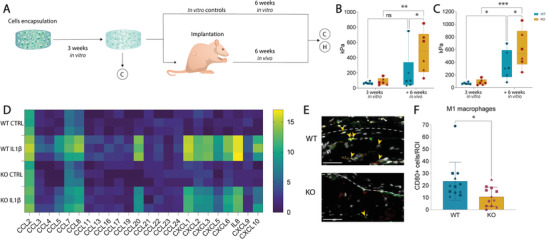
TAK1‐KO cells‐derived neocartilage develops into stiff matrix in vivo and recruits less M1 macrophages than WT‐derived one. A) Schematic of the experiment. WT and KO cells were encapsulated, samples were cultured for 3 weeks before subcutaneous implantation into nude rats and explanted or cultured in vitro after further 6 weeks. Sample diameter and thickness were 4 and 1 mm, respectively. C = compression, H = histology. B,C) Compressive modulus of in vivo (B) and in vitro (C) WT and KO samples at 3 and 9 weeks (two‐way ANOVA, 3 donors, n = 6). D) Gene expression analysis of main chemokines by multiplexing of WT and KO cells ± IL1β (n = 3). E) Immunofluorescence of macrophages infiltration in in vivo samples. External part of the construct above the dotted line, construct below. The pro‐inflammatory M1 macrophages were identified with double positivity for CD68 (pan‐macrophage marker) and CD80 (M1 marker). Red: CD68, green: CD80, grey: Hoechst. Orange arrows pointing to double positive cells for CD68 and CD80. Scale bar, 50 µm. F) CD68–CD80 double positive cells per region of interest (ROI) (Unpaired t test, 3 donors, n = 12). ns: non‐significant. Error bars represent SD. *p < 0.05, **p < 0.01, ***p < 0.001.
