Figure 3.

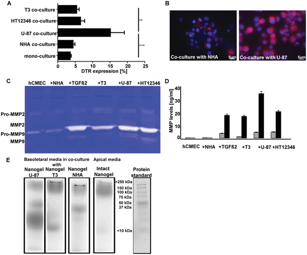
Enzymatic degradation of MMP‐substrate cross‐linked nanogels. A) DTR expression on hCMEC/D3 cells in different co‐cultures after 3 days estimated by flow cytometry (n = 5; one‐way ANOVA; Sidak's multiple comparisons test; **p < 0.01, ***p < 0.001). B) Visualization of membrane associated expression of DTR on hCMEC/D3 cells in co‐culture with NHA or U‐87 using fluorescence microscopy (DAPI: nucleus staining, 40‐fold magnification). C) MMP2 and MMP9 activities after 3 days in the media of mono‐culture, media of co‐culture with NHA, in media supplemented with TGFß2 (25 ng mL−1), in media of co‐culture with glioblastoma cell lines T3, U‐87, or HT12346 were detected by zymography. D) Quantification of MMP 2 (gray bars) and MMP9 (black bars) levels using ELISA (n = 5). E) Native gel electrophoresis /phosphor imager analysis of nanogel from media collected basolateral of the trans‐well, visualizing post transcytosis nanogel degradation products (90 min incubation with 1°MBq per well). The fourth lane visualizes the apical media collected from the co‐culture with U‐87 cells.
