
Figure 2.
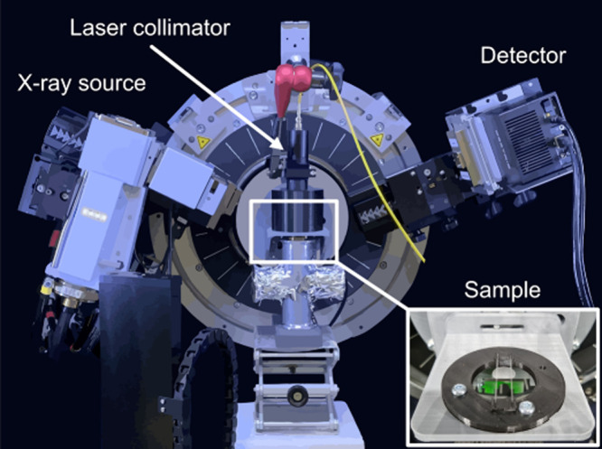
Schematic illustration of the setup used for the in situ XRD NIR laser experiments. The samples were placed on a support stage consisting of two glass capillaries (inserted picture), ensuring that the NIR beam did not heat any other elements than the sample. The NIR laser system’s collimator was placed 10 cm away from the sample at a 90° angle, illuminating the entire sample surface and ensuring that the flux was comparable between experiments.