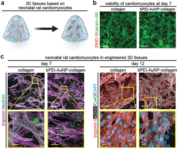
Figure 4.

bPEI‐AuNP‐collagen hydrogels support viability and subcellular organization of neonatal rat ventricular cardiomyocytes. a) Schematic illustration of generating engineered cardiac tissues containing neonatal rat ventricular cardiomyocytes. b) Representative images of neonatal rat ventricular cardiomyocytes cultured within hydrogels for 7 days stained with calcein‐AM (green, living cells) and ethidium homodimer‐1 (EthD‐1, red, dead cells). c) Examples of projections of confocal images of neonatal rat ventricular cardiomyocyte‐laden tissue constructs stained for the cardiomyocyte‐specific markers troponin I and sarcomeric‐α‐actinin (day 7 post‐fabrication) and connexin 43 (Cx43, day 12 post‐fabrication). DNA was visualized with DAPI. Scale bars: yellow: 5 µm and white: 25 µm.
