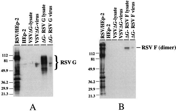
FIG. 3.
Western blot analysis of cell lysates and purified virions. (A) Proteins from uninfected and infected cell lysates were separated in a 12% polyacrylamide (SDS) gel and transferred to nitrocellulose. The blot was probed with a goat anti-RSV antiserum, and antibody binding was detected by ECL following binding with a horseradish conjugated-donkey anti-goat immunoglobulin G. The position of the heavily glycosylated RSV G is noted. Positions of molecular weight markers are noted on the left. (B) Proteins were separated in a 12% polyacrylamide (SDS) gel under nonreducing conditions. Samples were treated with SDS at room temperature prior to electrophoresis. Detection of the proteins bound by the goat anti-RSV antiserum is described above. The position of the RSV F 140-kDa dimer is noted, as are the positions of the molecular mass markers.

