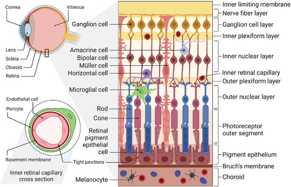
Figure 3.

Schematic presentation of the main anatomy of the ocular fundus. The ILM and BRB represent crucial barriers restricting the fundus drug delivery. The BRB is composed of two primary parts: the inner BRB, characterized by tight junctions between RECs, and the outer BRB, formed by tight junctions between RPE cells. Reproduced under the terms of the Creative Commons Attribution license 4.0 (CC‐BY) (https://creativecommons.org/licenses/by/4.0/).[ 29 ] Copyright 2023, The Authors, published by Elsevier.
