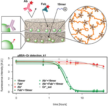
Figure 17.

Spatial separation of fluorescent microbeads into two detection levels by a hydrogel. The mesh size of the used hydrogel selected which molecules could penetrate by size exclusion. If a mixture of labeled antibody (Ab), fragment antigen binding (Fab), and oligonucleotide (19mer) was added, each molecule was detected in a different level and the antibody was excluded completely. The graph depicts the detection of the quencher labeled oligonucleotide, which proceeded in the bottom layer, and was achieved successfully in all cases were the 19mer was added (green lines). All negative controls retained their fluorescence intensity (red and grey lines). Adapted with permission.[ 148 ] Copyright 2019, American Chemical Society.
