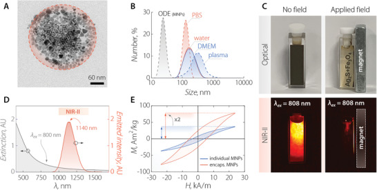
Figure 1.

A) TEM image of a single NIR‐MNC consisting of Ag2S NPs and Fe3O4 MNPs co‐encapsulated by phospholipids. MNPs of larger size form a packed corona in the proximity of the nanocapsule's surface, surrounding smaller Ag2S NPs. B) Comparison between the hydrodynamic size distribution obtained via DLS on colloidal dispersions of MNPs in octadecene (ODE) and NIR‐MNCs in distilled water, phosphate‐buffered saline PBS 1×, DMEM, and blood plasma. C) Optical (top) and NIR‐II (bottom) fluorescence images of a colloidal dispersion of NIR‐MNCs in the absence (left) and presence (right) of a magnetic field gradient created by a neodymium magnet. NIR‐MNCs are dragged towards the magnet so the solution becomes clear and luminescence is only generated in the vicinity of the magnet. D) Room‐temperature extinction (gray line) and emission (orange line) spectra of a colloidal dispersion of NIR‐MNCs. The emission spectrum was obtained under 800 nm optical excitation. E) Comparison between the AC magnetic hysteresis loops obtained at 100 kHz and 24 kA m−1 from individual (blue line) and encapsulated (orange line) MNPs dispersed in 1‐octadecene and in double‐distilled water, respectively.
