
Figure 6.
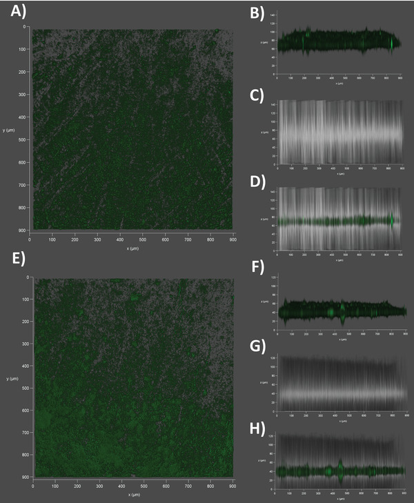
Confocal Laser‐Scanning Microscopy of the outer membrane vesicle (OMV)‐derived membrane model. A) Top view on an NBD‐PE‐stained (green) E. coli BL21 DE3 OMV‐coated filter support at dry state. B) Sideview on the same dry membrane using the fluorescence channel or C) the brightfield. D) Superimposition of fluorescence and brightfield channel. E) Top view on the same NBD‐PE‐stained and OMV‐coated filter support at wet state, after incubation for 30 min in PBS (pH7.4). F) Sideview on the membrane using the fluorescence channel or G) the brightfield. H) Superimposition of fluorescence and brightfield channel.