Figure 7.

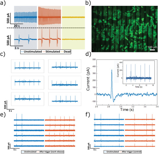
Printed MEA using silk hydrogel substrates and passivation for extracellular recording. a) Action potential recordings where initially cells are firing at a frequency of 0.3 Hz (blue), whereas addition of noradrenaline into the culture medium increased the beating frequency (red) and SDS starts to perforate and dissolve the cellular layer leading to stopping the beating of the cells (yellow). b) Fluorescence microscopy image of live/dead staining of HL‐1 cells growing on a silk MEA (scale bar 100 µm). c,d) Action potential recordings from HL‐1 cells using printed hydrogel electrode. e) Action potential recordings before and after the voltage trigger had locally released the printed noradrenaline. f) The voltage trigger's effect on HL‐1 cells cultured without noradrenaline under the electro‐responsive silk–carbon layer.
