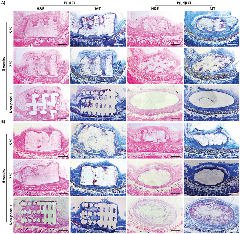
Figure 6.

Immune response to dual‐porosity scaffolds in vivo. Histological staining of nonporous and dual‐porosity scaffolds prepared from 7% and 5% copolymer gels of P(l)LCL and P(l,d)LCL after 3 and 6 weeks of implantation on a rat subcutaneous model. Samples were stained for hematoxylin and eosin‐Y (H&E) and for Masson's trichrome (MT). Scale bar in all pictures is 1 mm.
