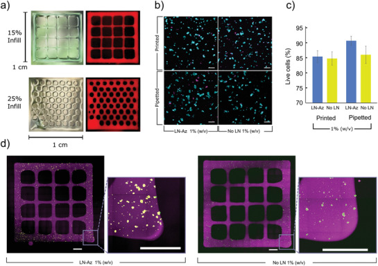
Figure 7.

a) 3D bioprinted structures based on the HA:PEG‐LN hydrogels at a concentration of 1% (w/v). Hydrogels (red) were dyed with Cy5 and illuminated using a white light source. b) Live (cyan)/Dead (magenta) staining of SH‐SY5Y cells 24 h after bioprinting. c) SH‐SY5Y cell viability 24 h after bioprinting or pipetting when encapsulated in either HA:PEG (without LN) and HA:PEG‐LN hydrogels at a concentration of 1% (w/v), N = 4 bioprinted/pipetted hydrogels were examined for each condition, error bars are standard deviation, statistics: one‐way ANOVA with Tukey's HSD, n.s. = not significant (p > 0.05). d) SH‐SY5Y cell bioprinted into grid structures (purple) of Cy5‐labeled HA:PEG‐LN and HA:PEG, respectively, at a hydrogel concentration of 1% (w/v) and imaged using tiled confocal microscopy 24 h after bioprinting. Encapsulated SH‐SY5Y were stained using Live (cyan)/Dead (magenta) staining. Inset square indicates a magnified portion. Scale bars are 1000 µm.
