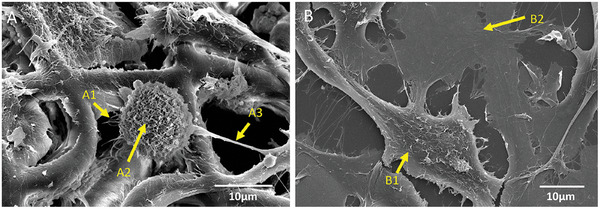
Figure 3.

Detailed SEM micrographs of the U251/HUVEC Co‐culture. A) Co‐cultured cells on 3D micro‐vessels‐like structures ([A–1] indicates minor extensions of the cells, [A–2] indicates the microvilli on the surface of the cells, and [A–3] indicates the major extensions). B) Co‐cultured cells on 2D pedestals ([B–1] indicates U251 cells, [B–2] points at HUVECs).
