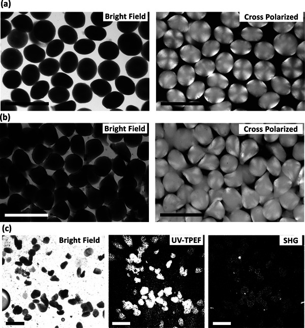
Figure 5.

Microscopy images of the crystalline monoclonal antibody (mAb) laden alginate hydrogel particles. a) Bright‐field micrograph (left) and cross‐polarized microscopy (right) at 100 mg mL−1 particle loading. b) Bright‐field micrograph (left) and cross‐polarized microscopy (right) at 350 mg mL−1 particle loading. The cross‐polarized microscopy images of the crystalline mAb‐laden hydrogel particles indicated the crystals loaded within the hydrogel particles. c) Bright‐field microscopy (left), ultraviolet two‐photon excited fluorescence (center), and second‐order nonlinear optical imaging of chiral crystals (SONICC, right) imaging of chiral crystals at 250 mg mL−1 particle loading. Scale bars are 400 µm.
