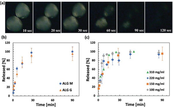
Figure 7.

Dissolution and release of the anti‐PD‐1 antibody from the alginate hydrogel particles in simulated body fluid (SBF) at 37 °C. a) Time lapse of cross‐polarized microscopy imaging of the hydrogel particles immersed in SBF which indicates that the antibody crystals are present (bright) at t = 10 s and are dissolved (dark) at t = 120 s. The sample was prepared at 200 mg mL−1 particle loading using ALG M. b) Release profiles of monoclonal antibody (mAb) from guluronic rich (ALG G) and mannuronic rich (ALG M) hydrogel particles at 150 mg mL−1 particle loading. c) Release profiles of the mAb from particles with varying particle loadings indicating that the mAb is completely released from the particles within minutes. Samples in triplicate.
