
Figure 4.
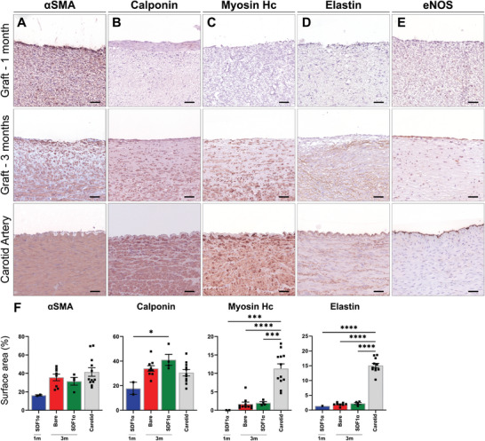
Immunohistochemistry on vascular markers in representative transverse sections of the center part of the grafts, at 1 month (n = 2), 3 months (Bare n = 9, SDF1α n = 4), and native carotids (n = 12) as controls. Stained with Novared for A) smooth muscle marker αSMA, B) early contractile marker Calponin, C) mature contractile marker Myosin Heavy Chain, D) Elastin and E) endothelial marker eNOS. With measured positive surface area of whole cross sections taken of separate parts of the grafts. Electrospun fibers are visible in white. Scale bars 50 µm. Data are shown as mean ± SEM. *p < 0.05, ***p < 0.001, ****p < 0.0001.