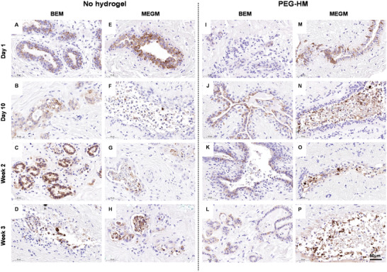
Figure 4.

Epithelial staining of normal breast (NB‐2) explant cultures. Tissue explant cultures of normal breast tissue were cultured embedded in PEG‐HM in BEM or MEGM medium. Control tissues were cultured in parallel without prior embedding. To confirm the presence of epithelial cells in the mammary tissues, 4 µm thick sections of explant cultures were stained for Cytokeratin 8/18. Representative brightfield images of epithelial glandular structures are shown of NB‐2 explants cultured in A–D) BEM medium without hydrogel embedding, I–L) embedded in hydrogel, E–H) as well as cultured in MEGM medium without hydrogel embedding and M–P) embedded in hydrogel. Scale bar: 50 µm.
