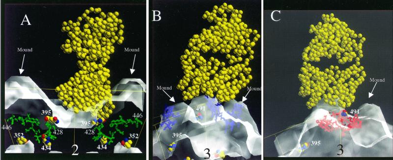
FIG. 7.
Side-view models of ADV-GVP2 particles complexed with IgG Fab fragment (depicted as van der Waals balls). The position of the icosahedral threefold related mounds and residues involved in host range and pathogenicity (VP2:352, VP2:395, VP2:434) (14, 31) is noted. (A) Fab fragment (yellow) docked with VP2:428-446 (green) in a side view perpendicular to an icosahedral twofold axis. (B) Fab fragment (yellow) docked with VP2:455-470 (blue) in a side view perpendicular to an icosahedral threefold axis. (C) Fab fragment (yellow) docked with VP2:487-501 (red) in a side view perpendicular to an icosahedral threefold axis.

