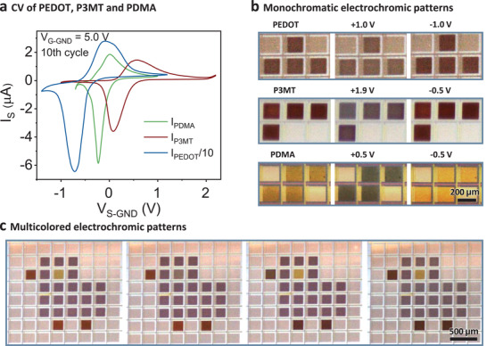
Figure 4.

Site‐selective deposition and color switch of multicolored electrochromic patterns. a) CV of PEDOT, P3MT, and PDMA on individual active‐matrix pixels. The V G‐GND is kept as 5 V during the source bias scan. b) Optical microscopy images of PEDOT, P3MT, and PDMA patterns in the as‐deposited, oxidation and reduction states, respectively. For the oxidation and reduction states, the applied V S‐GND are marked on top of the microscopy images. c) PEDOT, P3MT, and PDMA are sequentially deposited on the same active‐matrix and the pixels within the pattern can be changed between different electrochromic colors switched by the a‐IGZO TFT active‐matrix. The PDMA eye displays color changes between yellow and green, the P3MT beak and legs show color changes between red and dark blue, and the PEDOT body shows colors between dark blue and light blue.
