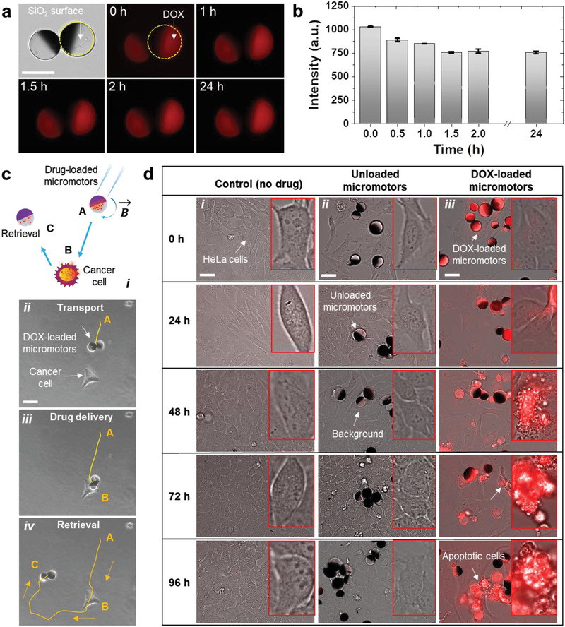
Figure 5.

Study of DOX‐loaded micromotors functionality. a) Fluorescence images of DOX‐loaded micromotors over 24 h, Scale bar: 20 µm. b) Fluorescence intensity of the drug‐loaded micromotors. c) Schematic showing the transport of drug‐loaded micromotors from position A to the target HeLa cell (position B). After releasing the drug, the micromotor can be retrieved by applying an inverse rotating magnetic field, and be transported to the desired location (position C). Optical images show the complete trajectory indicating i) transport, ii) drug‐release, and iii) retrieval of a micromotor. Scale bar: 20 µm. d) Time‐lapse fluorescence images showing DOX diffusion by the transported micromotors onto the 2D cell culture. Images were taken at an excitation wavelength of 470 nm and an emission wavelength of 580 nm. Overlaid BF and fluorescence images captured from 0 to 96 h for each group: i) control sample, ii) with unloaded micromotors, and iii) with drug‐loaded micromotors. Scale bar: 20 µm. HeLa cells were cultured in a coverslip in a cell culture medium and grown for 3 d in the incubator at 37 °C and 5% CO2, cell count = 5 × 10⁴ for each sample.
