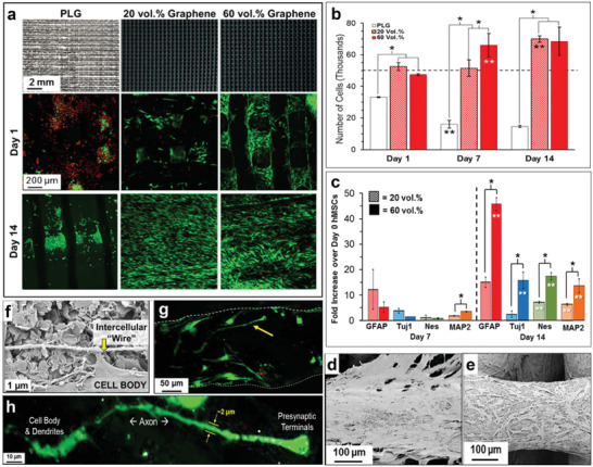
Figure 5.

Example of osteogenic behavior of graphene scaffolds. a) Top row: Photographs of 3D printed scaffolds of PLG materials with 0%, 20%, and 60% graphene (volume %); Scanning laser confocal 3D reconstruction projection of live (green) and dead (red) human mesenchymal stem cells (hMSCs) cultured at day 1 (middle row) and day 14 (bottom row) on the scaffolds. b) Number of hMSCs present on the scaffolds at day 1, 7, and 14, suggesting superior cell proliferation on the scaffolds with higher concentration of graphene. c) Neurogenic relevant gene expression of the cells on the graphene scaffolds at day 7 and 14, showing better gene expression with higher content of graphene. d,e) SEM of hMSCs on the 20% and 60% graphene loaded scaffolds, respectively. f) High magnification SEM of hMSCs cell on 60% graphene scaffold at day 7, showing hMSCs connecting via a long “intercellular” wire. g) Scanning laser confocal 3D reconstruction of live (green) and dead (red) hMSCs cells on day 14 for 60% graphene scaffold. h) High magnification image of the cell indicated by yellow arrow in (f) showing the detailed features. Reproduced with permission.[ 95 ] Copyright 2015, American Chemical Society.
