
Figure 2.
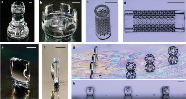
3D microstructuring of fused silica glass using DLW. a,b) Exemplary microrook with a height of ≈2 mm (scale bars: (b): 200 µm). c,d) Microfilter element with a pore size of 55 µm (scale bars: 500 µm). e) Fused silica glass upright lens (scale bar: 180 µm). f) Side view of the standing lens of (e) (scale bar: 190 µm). g) Three fused silica upright microlenses directly printed on a silicon substrate with three Wigner–Seitz cells printed in IP‐Q (scale bar: 400 µm). h) Front view through the three fused silica glass microlenses in (g) showing the cells at different distances (scale bar: 360 µm).