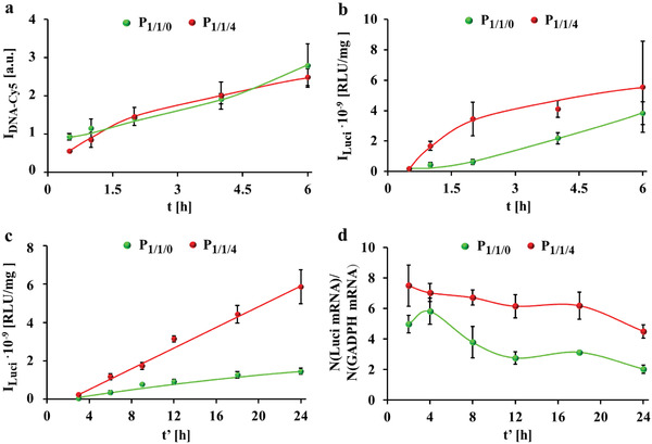
Figure 1.

Cellular uptake, luciferase expression, and mRNA transcription. a) Association of P 1/1/0 and P 1/1/4 polyplexes prepared from Cy5‐labelled DNA (DNACy5) to HeLa cells in terms of Cy5 fluorescence I DNA‐Cy5 per cell as measured by flow cytometer. Cells were incubated with polyplexes at 1.4 µg mL−1 DNA (containing 40 ng mL−1 DNACy5) in serum free culture medium for t = 0.5–6 h and measured immediately. b) Luciferase expression of cells after exposure to polyplexes prepared from luciferase‐encoding plasmid (pLuci) for different incubation times t. Cells were incubated with polyplexes at 1.4 µg mL−1 pLuci in serum free medium for t = 0.5–6 h, followed by further culture in fresh 10% FBS supplemented cell culture medium without polyplexes for t′ = 24 h. The luciferase luminescence I Luci from cells was detected by a luminescence detection system. Luminescence is given in relative luminescence units (RLU) per amount of total proteins in RLU/mg. c) Luciferase expression I Luci after different culture time t′. Cells were incubated with polyplexes at 1.4 µg mL−1 DNA in serum free medium for t = 3 h, followed by further culture in fresh 10% FBS supplemented cell culture medium for t″ = 3–24 h. d) mRNA transcription upon exposure of cells to polyplexes at 1.4 µg mL−1 DNA in serum free medium for t = 3 h, followed by further culture in fresh 10% FBS supplemented cell culture medium for t' = 2–24 h. mRNA transcription is displayed in terms of the relative luciferase mRNA transcription number normalized to the house keeping gene GADPH, that is, detected number of luciferase mRNA/detected number of GADPH mRNA.
