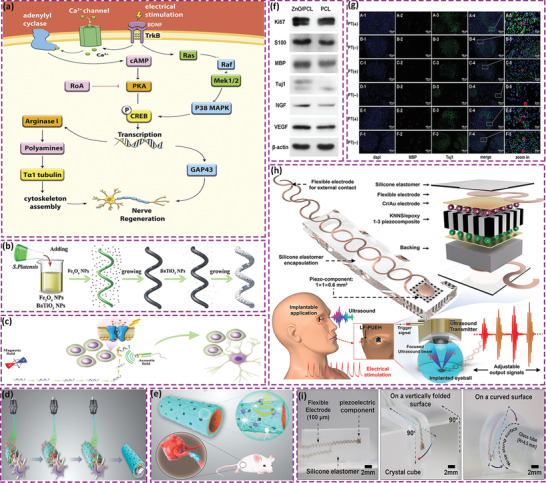
Figure 9.

a) Regulation of electrical stimulation on metabolic pathways in neuronal cells. Reproduced with permission.[ 158a ] Copyright 2020, Elsevier. b) Schematic of the fabrication of Spirulina platensis@Fe3O4@tBaTiO3 micromotor. c) Magnetism‐driven movement to the targeted neural stem‐like cell and ultrasonic‐induced cell differentiation. Reproduced with permission.[ 159 ] Copyright 2020, Wiley‐VCH. d) Preparation of ZnO loaded PCL conduit via 3D injectable multilayer biofabrication. e) Mechanical stimulation induced piezoelectric effects. f) Western blotting results of Ki67, S100, MBP, Tuj1, NGF, and VEGF in Schwann cells on ZnO/PCL and PCL scaffolds. g) MBP and Tuj1 triple immunostaining image of sciatic nerves acquired from the ZnO/PCL, PCL, and autograft groups: (A1–A5, B1–B5) ZnO/PCL conduit; (C1–C5, D1–D5) PCL conduit; (E1–E5, F1–F5) autograft. PT (+): physical therapy group. PT (‐): nonphysical therapy group. Reproduced with permission.[ 104 ] Copyright 2020, Wiley‐VCH. h) Schematic of key components of a device which utilizes lead‐free piezoelectric composite as the core component, wavy‐structure‐based flexible electrodes as external contact, and a silicone elastomer as encapsulation. The device provides adjustable electrical outputs driven by ultrasound for electrical stimulation. i) Images of device to demonstrate mechanical properties. Reproduced with permission.[ 161 ] Copyright 2019, Wiley‐VCH.
