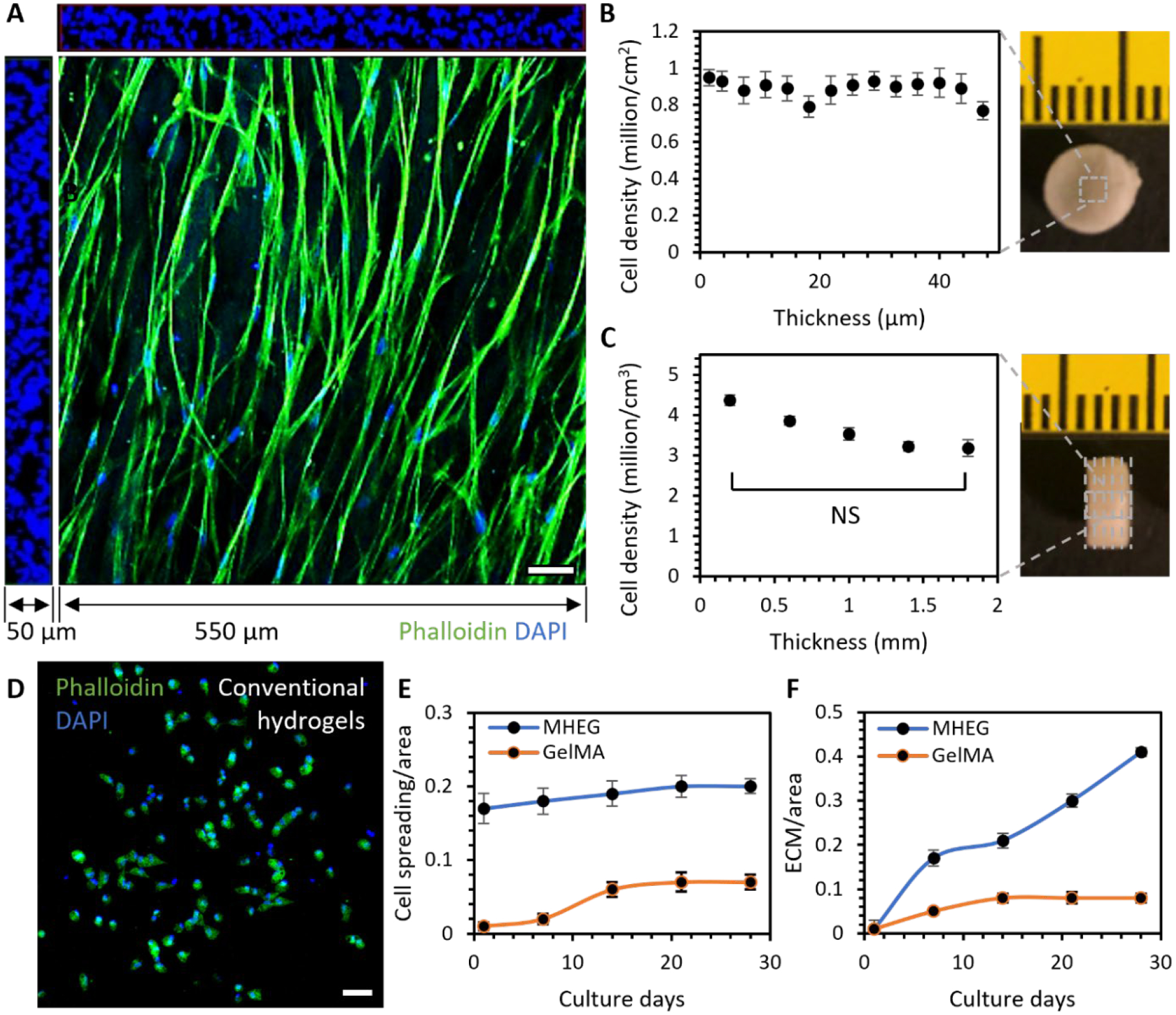
Figure 6. ECM-like cell functional property.

A. The 3D rendering of confocal stacks with dimensions of 550 × 550 × 50 μm showed that cells formed an interconnected 3D meshwork in MHEG. B. Cell density in each 2D stack throughout the 50 μm thickness. C. Cell density in each 3D volume at different depths of a sample with a thickness of 2 mm. D. Cells were normally isolated inside conventional GelMA hydrogels. E. Comparison of cell spreading area/image area in MHEG and GelMA with increasing culture time. F. Comparison of newly deposited ECM area/image area in MHEG and GelMA with increasing culture time. n = cells in a volume of 3 × 550 μm × 550 μm × 50 μm, two tailed student T test. Scale bar: 50 μm.
