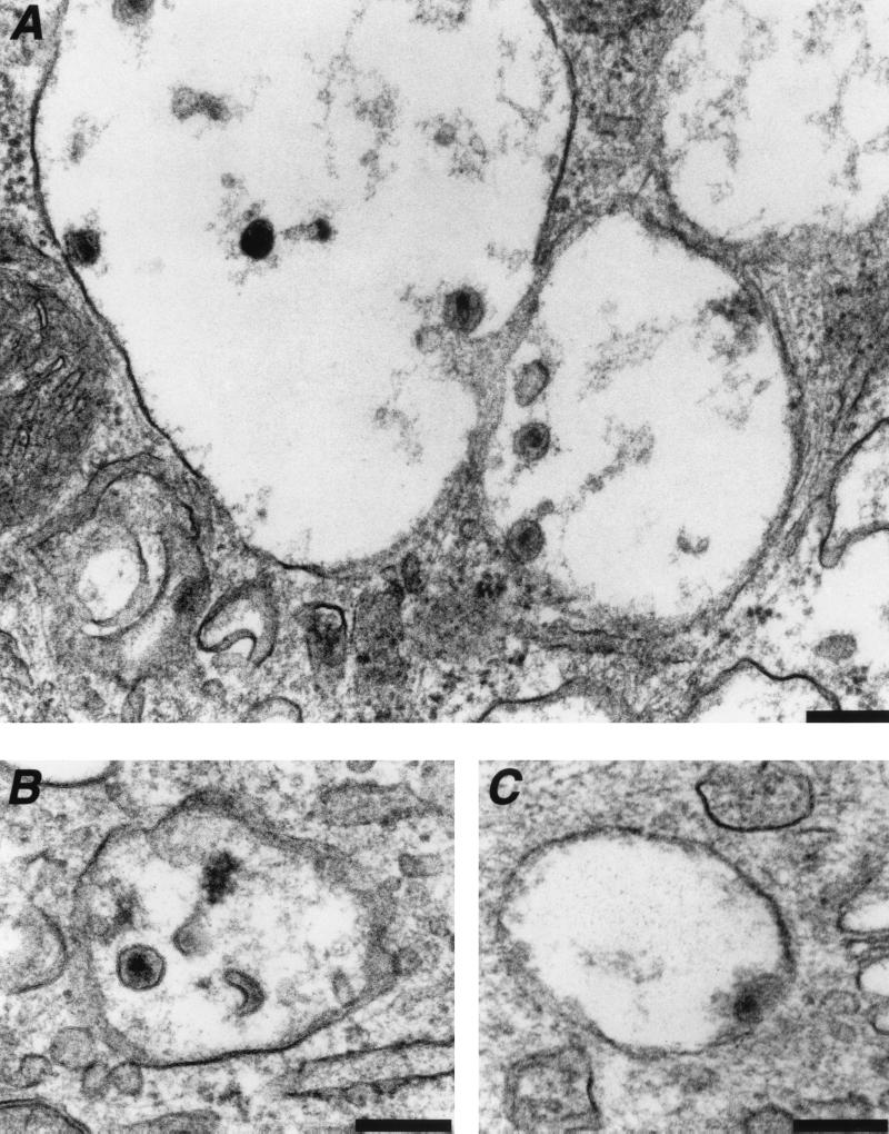
FIG. 3.
Fate of HIV-1 virions internalized in intracellular vesicles. Macrophages were exposed to the R5-tropic HIVNLAD8 for 30 to 45 min and processed for electron microscopy. (A and B) Intact or degraded HIV-1 virions present in the lumen or bound to the membrane of intracellular vesicles. (C) Fusion between viral and vesicular membranes. Data are representative of three independent experiments. Bar = 0.2 μm.

