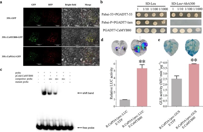
Figure 6.
CaMYB80 binds to the promoter of CaPOA1. (a) Subcellular localization; scale bars = 10 μm. (b) Results of the Y1H experiment. (c) EMSA, from left to right: 6-FAM labeled prope; CaMYB80 and 6-FAM labeled probe; CaMYB80, 6-FAM labeled probe and 10× unlabeled probe; CaMYB80, 6-FAM labeled probe and 20× unlabeled probe; CaMYB80, 6-FAM labeled probe and 50× unlabeled probe; CaMYB80 and 6-FAM labeled mutant probe. (d) LUC analysis results. (e) GUS enzyme activity results. Asterisks indicate significant difference (*P < 0.05; **P < 0.01).

