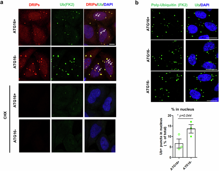
Extended Data Fig. 5. Puromycin-induced misfolded protein inclusions are found in the nucleus in ATG16L1 KO cells.
a, ATG16L1 WT (ATG16+) and KO (ATG16-) HeLa cells were treated with O-propargyl-puromycin (OP-Puro, DRIPs) with or without cycloheximide (CHX, 50 µg/ml) as a control for 2 h. Cells were fixed and OP-Puro and ubiquitin-positive structures were visualized using confocal microscopy. Arrow indicates DRIPs in the nucleus. Scale bar, 10 µm. (Representative images from 3 biological repeats) b, ATG16L1 WT and KO HeLa cells were treated with puromycin for 4 h. Cells were fixed and ubiquitin-positive structures were visualized using confocal microscopy. Total area of ubiquitin-positive structures (Ub+) in nucleus and entire cell was quantified. Scale bar, 20 µm. Values are mean ± S.E.M. (n = 3 independent experiments; * p < 0.05 vs. WT cells; two-tailed paired t-test). Source numerical data are available in source data.

