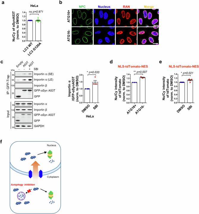
Extended Data Fig. 7. Autophagy compromise causes bulk proteins ‘overflow’.
a, Quantification of the ratio of nucleus/cytosol-localized αSyn A53T upon either LC3 wild-type (LC3 WT) or non-lipidated form (LC3 G120A) overexpression, by immunostaining. HeLa cells were transfected pHM6-αSyn A53T (HA tag) with either GFP-LC3 wild-type (WT) or GFP-LC3 G120A mutant and then fixed for immunostaining using HA antibody to confirm the localization of αSyn A53T (Value are mean ± S.E.M, n = 6 biological independent experiments; ns = not significant vs. LC3 WT; two-tailed paired t-test). b, HeLa/ATG16L1 wild-type and null cells were fixed and labelled for NPC (nuclear pore complex, green), RAN (Ras-related nuclear protein, red), and DAPI (nucleus, blue). This experiment was performed once simply to confirm an intact NPC in ATG16L1 WT and KO cells. Scale bar, 20 μm. c, Representative blots showing the binding of αSyn A53T with importin α upon autophagy inhibition (SBI, 5 uM) in HeLa cells. Cells were transfected with either GFP-empty or GFP-αSyn A53T and then cells were treated with SBI. Then, immunoprecipitates obtained using GFP-trap were processed for immunoblotting to detect importin α and importin β. (Right graph) Quantification shows the amount of importin α bound GFP-αSyn A53T (Value are mean ± S.E.M; n = 4 biological independent experiments; * p < 0.05 vs. DMSO; two-tailed paired t-test). d and e, Cells were transfected with NLS–tdTomato-NES which is a shuttling reporter containing both an NLS and an NES fused to tdTomato. d, Quantification of a ratio of nucleus/cytosol-localized tdTomato intensity in HeLa/ATG16L1 wild-type (ATG16+) and null cells (ATG16-) (Value are mean ± S.E.M, n = 3 independent experiments; ** p < 0.01 vs. ATG16 + ; two-tailed paired t-test). e, Quantification of a ratio of nucleus/cytosol-localized tdTomato intensity upon SBI treatment for 15 h in HeLa cells. (Value are mean ± S.E.M, n = 5 biological independent experiments; * p < 0.05 vs. DMSO; two-tailed paired t-test) f, Schematic representation shows autophagy compromise leads to an “overflow” of bulk proteins (likely autophagy substrates) into the nucleus for degradation by nuclear proteasomes. Source numerical data and unprocessed blots are available in source data.

