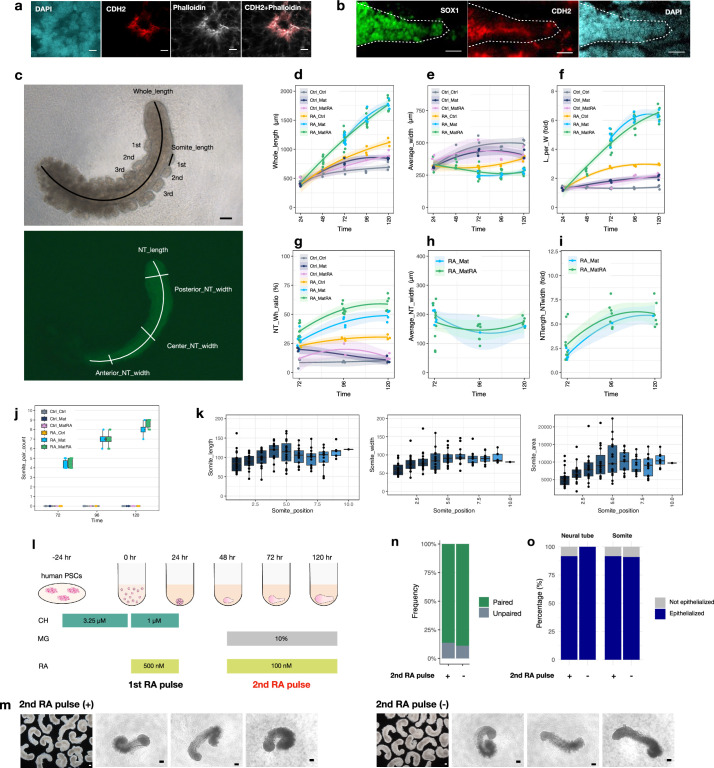
Extended Data Fig. 5. Morphological properties of human conventional and RA-gastruloids.
a, Immunostaining of N-cadherin (CDH2) and phalloidin in somites in an RA-gastruloid. Phalloidin-stained F-actin and CDH2 were co-localized and highly concentrated at the apical surface of somites. Scale bar, 10 µm. b, Immunostaining of N-cadherin (CDH2) and SOX1 in the neural tube in an RA-gastruloid. Scale bar, 100 µm. c, (Top) Bright-field of a human RA-gastruloid. The whole length was measured as the length of a line along the centre of the body. Each somite length was measured from the posterior end. (Bottom) SOX2-mCit view of the top picture. Neural tube length (NT_length) was measured as the continuous SOX2+ area. The width of neural tubes was measured and averaged over several positions (10%, 50%, 90% along the full length of the structure). Scale bar, 100 µm. d-l, Morphometric measurements of gastruloids which originated from 5,000 cells, as a function of time. Ctrl, no treatment controt; RA, Retinoic acid; Mat, 5% Matrigel; MatRA, Matrigel + RA. Left and right part of each text label indicates the conditions at 0–24 h and at 48–120 h, respectively. For example, Ctrl_Ctrl indicates no treatment for both 0–24 h and 48–120 h. N = ≥ 3 for each time point and condition. d, Whole length (µm) of gastruloids. e, Average width (µm) of gastruloids. f, Ratio (%) of whole length to average width. g, Ratio (%) of length of neural area to the whole length. h, Average neural tube width (µm). i, Ratio (%) of neural tube length-to-width. j, Number of somites observed as a function of time. k, Length, width, and area of somites as a function of position. N = 16 RA-gastruloids. l, Schematic of RA-gastruloid induction protocol, highlighting the first vs. second RA pulse. m, Bright-field images of human RA-gastruloids induced with (left column) vs. without (right column) inclusion of the second RA pulse. Scale bars = 100 µm. n, Frequency of paired somites in RA-gastruloids with vs. without inclusion of the second RA pulse. Somites with areas within 30% of one another were classified as "paired somites". This comparison was made for 3 randomly chosen putative somite pairs within each gastruloid. A gastruloid was subsequently designated as "paired gastruloid" if at least 2 out of 3 putative somite pairs were classified as "paired somites". N = 13/14 (92.9%) and N = 11/12 (91.7%) for RA-gastruloids with vs. without inclusion of the second RA pulse, respectively. o, Frequency of neural tube (left) and somite (right) epithelialization with vs. without inclusion of the second RA pulse, respectively. Epithelization was defined by the accumulation of phalloidin staining at the apical side of the structures upon immunostaining. The percentages indicate the frequency of gastruloids with epithelialized somite or neural tube. N = 11 and N = 10 for RA-gastruloids with vs. without inclusion of the second RA pulse, respectively.

