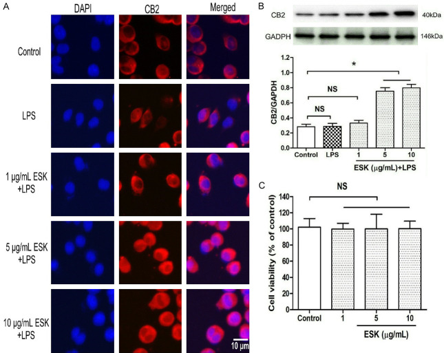
Figure 2.
Esketamine upregulated CB2 protein expression in microglial cells. The grouping and treatments were the same as in Figure 1, and immunocytochemistry staining and western blot analysis were performed to assess CB2 expression. Then, the cells were assigned into four groups, including the normal cultured control and three concentrations of esketamine (ESK) groups. After 24 h of treatment, the cell injury was assessed by using the MTT assay. A. CB2 receptor staining (magnification: 40×10). B. CB2 expression level (n = 4). C. Microglial viability (n = 10). *: P < 0.05; NS: no significance.

