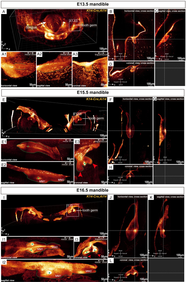
Figure 2.
MiniTESOS clearing-based deep three-dimensional imaging enables imaging of tooth germ morphology at embryonic stages with high resolution. (A) A three-dimensional view of the mandible from K14-Cre;Ai14 mice at E13.5; scale bars: 100 μm. (A1-A3) The mouse molar tooth germ at E13.5 is shown in three dimensions. (A1) represents the horizontal view. (A2) represents the sagittal view. (A3) represents the coronal view; scale bars: 50 μm. The green dashed lines circle the outline of the tooth germ. (B-D) MM1 is shown in the horizontal, sagittal, and coronal cross sections, respectively, at E13.5. The white horizontal dashed lines indicate the sections on the same coronal plane, whereas the white vertical dashed lines indicate the sections on the same sagittal plane; scale bars: 100 μm. (E) A three-dimensional view of the mandible of K14-Cre;Ai14 mice at E15.5; scale bars: 100 μm. (E1-E3) The mouse molar tooth germ at E15.5 is shown in three dimensions. (E1) represents the horizontal view, (E2) represents the sagittal view, and (E3) represents the coronal view; scale bars: 50 μm. The green dashed lines circle the outline of the tooth germ. Red arrowheads show the cervical loop. The green asterisk shows the primary enamel knot; scale bars: 100 μm. (F-H) Horizontal, sagittal, and coronal cross-sections of MM1 at E15.5. The white horizontal dashed lines indicate sections on the same coronal plane, while the white vertical dashed lines indicate sections on the same sagittal plane; scale bars: 100 μm. (I) Three-dimensional view of the mandible from K14-Cre;Ai14 mice at E16.5; scale bars: 100 μm. (I1-I3) Three-dimensional views of the mouse molar tooth germ at E16.5 are shown in three dimensions. (I1) shows the horizontal view, (I2) shows the sagittal view, and (I3) shows the coronal view; scale bars: 50 μm. Green dashed lines outline the tooth germ. Red arrowheads indicate the cervical loop. Green asterisks denote the second enamel knots. (J-L) Horizontal, sagittal, and coronal cross sections of MM1 at E16.5. The white horizontal dashed lines indicate sections on the same coronal plane, and the white vertical dashed lines indicate sections on the same sagittal plane. MM1, mandibular first molar; MM2, mandibular second molar; scale bars: 100 μm; n = 3 per group.

