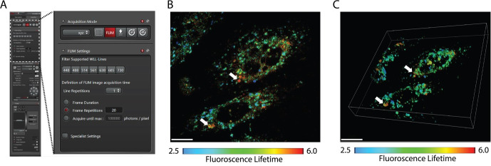
Figure 1. Illustration of Raw 3D FLIM image.
A. Settings for acquiring FLIM image using Leica Stellaris 8 confocal. B. Micrograph of 2D plane of CHO M21K F86L cells acquired using settings as in A. C. Micrograph of 3D FLIM image of cell as in B. White arrows point toward aggregates. Images are at 63× magnification and were acquired using Leica Stellaris 8 FALCON. Scale bar: 20 µm. Lifetime range: 2.5–6.0.

