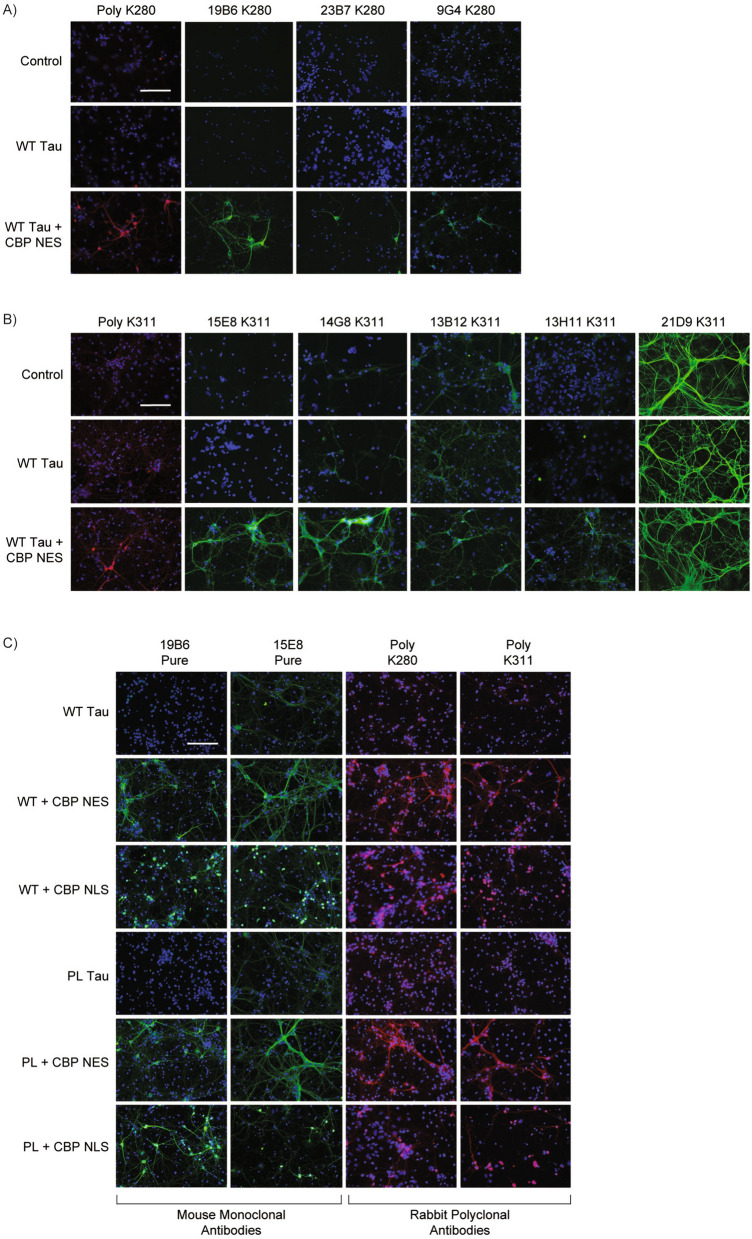
Fig. 3.
Immunocytochemistry validation of ac-tau monoclonal antibodies in primary neurons. Primary mouse cortical neurons were transduced for five days with lentiviruses for empty vector control, wild-type human tau, or wild-type tau and CBP-CD-NES. A Immunocytochemistry of neurons with rabbit polyclonal ac-K280 (RFP) and small-scale purification of 19B6, 23B7, and 9G4 (GFP). B Immunocytochemistry of neurons with rabbit polyclonal ac-K311 (RFP) and small-scale purification of 15E8, 14G8, 13B12, 13H11, and 21D9 (GFP). C Immunocytochemistry of large-scale purified 19B6 and 15E8 (GFP) and polyclonal ac-K280 and ac-K311 (RFP) after lentiviral transduction with wild-type or P301L human tau and CBP-CD-NES or CBP-CD-NLS. Scale bars, 125 μm. DAPI in blue

