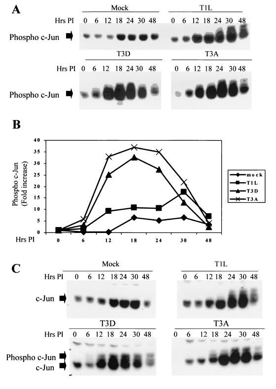
FIG. 2.
c-Jun is activated following infection with reovirus. Cells were infected with different strains of reovirus (MOI, 100) and were harvested at various times p.i. (A and C) Extracts were standardized for protein concentration, using an anti-actin antibody, and equal amounts of protein were separated by SDS-PAGE and probed with antibodies directed against phosphorylated (A) or total (C) c-Jun. Bands corresponding to phosphorylated and unphosphorylated c-Jun are shown. The gels are representative of at least two independent experiments. (B) Graphical representation of the Western blot shown in panel A, showing the fold increase in the levels of phophorylated c-Jun over time.

