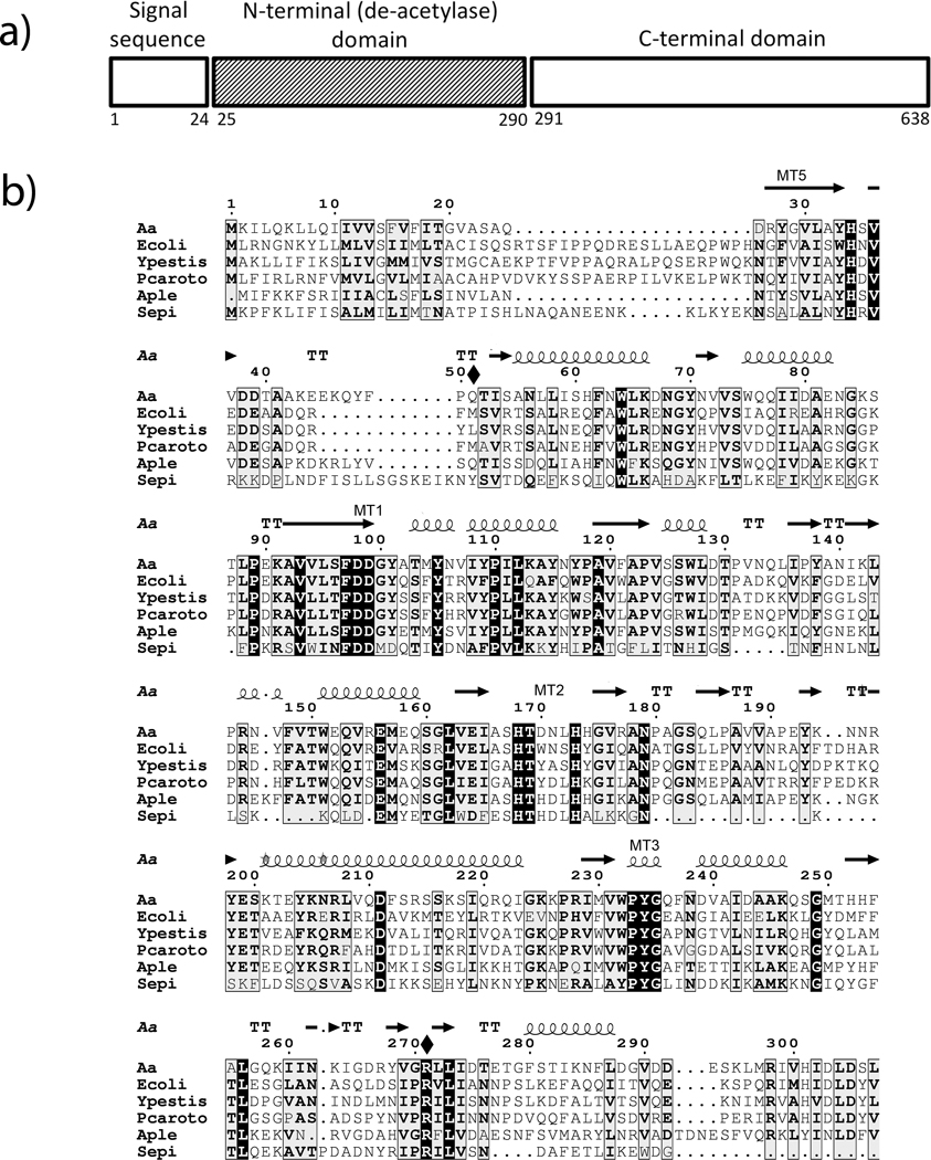
Figure 1.
Sequence homology among PgaB proteins. a) A schematic of the AaPgaB protein showing the various domains in the sequence. b) Sequence alignment of the N-terminal domain of PgaB proteins in A. actinomycetemcomitans (Aa),. E. coli (Ecoli), Y. pestis (Ypestis), P. coratovorum (Pcorato), A. pleuropneumoniae (Aple), and S. epidermidis (Sepi). The secondary structures as deduced from the AaPgaBN structure, the two mutated residues (shown with a solid diamond) and the conserved motifs are also shown above the sequence. Numbering is based on the A. actinomycetemcomitans sequence.

