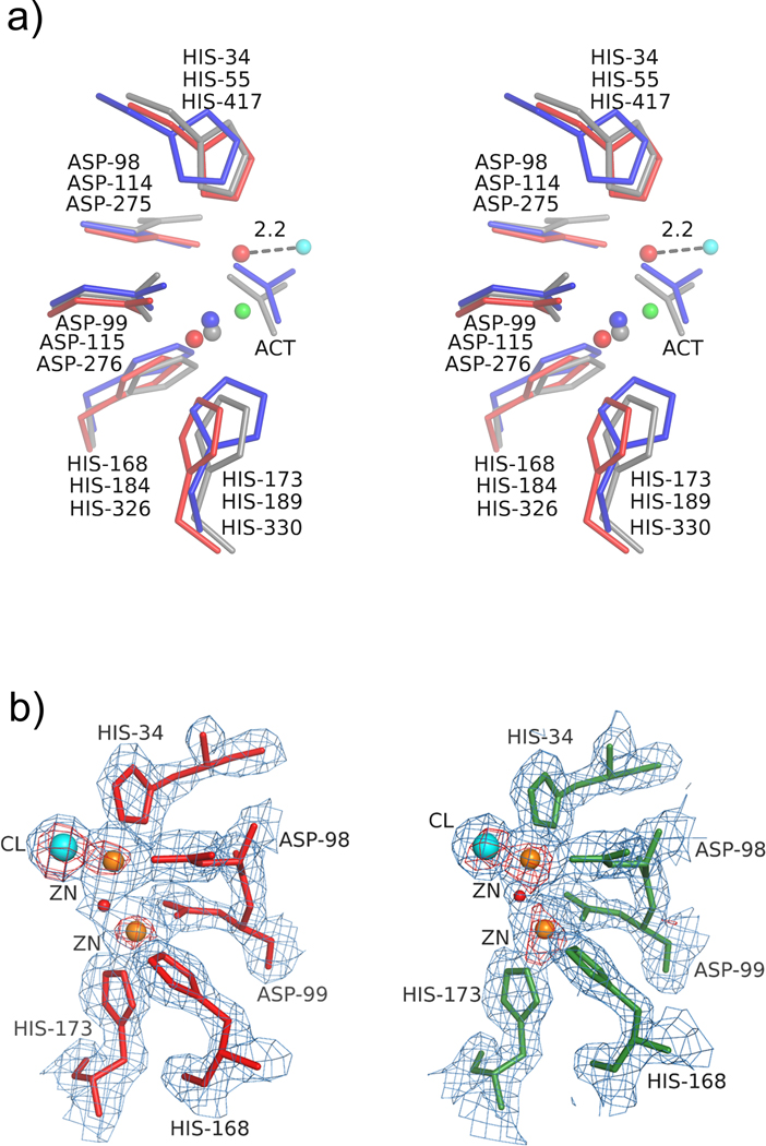
Figure 3.
Stereoview of the metal binding site in AaPgaBN. a) The critical residues in three de-N-acetylases, AaPgaBN (red, chain A), EcPgaB (grey) and SpPgdA (blue) are superposed; b) The anomalous difference density map (red contours) superposed with the 2Fo-Fc map (light blue) around the active site (Motifs MT1 and MT2) in chains A (left) and B (right) showing the metal ions (brown) and chloride (aqua). The nucleophilic water molecule is shown as a red sphere.

